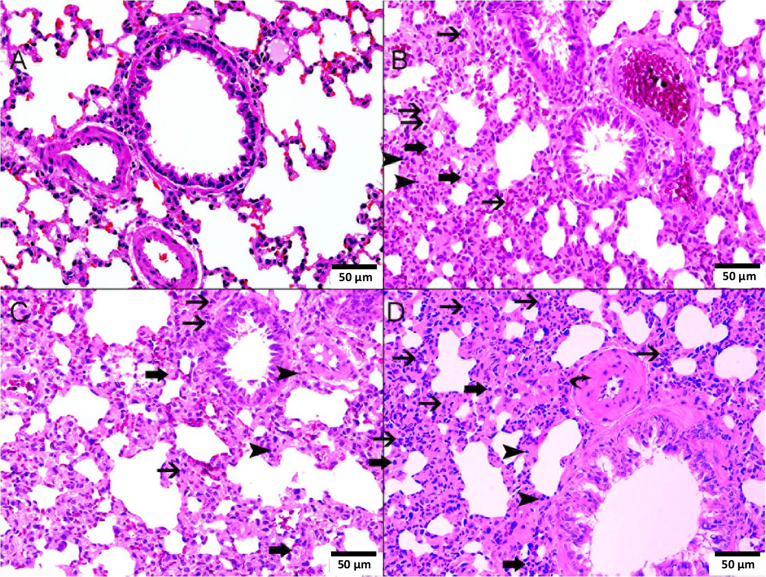
Fig. 6. A-D: representative light microscopy sections of H and E stained lung tissues of Wistar rats intratracheally instilled with either saline (control) or cerium oxide nanoparticles (CeO2 NPs) with or without cisplatin (CP) administration. A: representative lung sections obtained from saline-treated rats showing normal lung architecture and histology. B: representative lung sections obtained from CP + saline-treated rats showing the presence of moderate widening of interalveolar interstitial spaces with mixed inflammatory cells consisting of neutrophil polymorphs (thin arrow), lymphocytes (arrowhead) and macrophages (thick arrow). C: representative lung sections obtained from CeO2 NPs-treated rats showing the presence of foci of mild widening of interalveolar interstitial spaces with mixed inflammatory cells consisting of neutrophil polymorphs (thin arrow), lymphocytes (arrowhead) and macrophages (thick arrow). D: representative lung sections obtained from CP + CeO2 NPs-treated rats showing the presence severe widening of interalveolar interstitial spaces with mixed inflammatory cells consisting of neutrophil polymorphs (thin arrow), lymphocytes (arrowhead) and macrophages (thick arrow).